Symblepharon
Jump To
A symblepharon is a fibrous tract that connects bulbar conjunctiva to conjunctiva on the eyelid. It is expected after ulceration and exposure of subepithelial connective tissue. It is a pathologic condition and can be categorized as follows: Symblepharon, Normal Fornix and Symblepharon, Upper Fornix.


When do symblepharon occur?
Symblepharon results from an abnormal healing process after injury to the conjunctiva. Regardless of what causes the injury, the loss of epithelial cells from the bulbar and palpebral conjunctiva allows an abnormal adhesion to form between the bulbar and palpebral conjunctiva. Potential causes are listed below:
- Cicatricial pemphigoid: auto-immune disease which affects mucus membranes such as the mouth and oral pharynx, conjunctiva, nares and genitalia.
- Atopic keratoconjunctivitis: a hypersensitivity to environmental allergies including asthma, rhinitis, dermatitis and eczema.
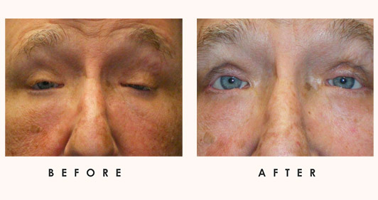
- Trichiasis: a lid abnormality in which eyelashes are misdirected towards the eyeball. These misdirected lashes might often be the lead to of scarring.
- Toxic epidermal necrosis: a potentially life-threatening disorder which is commonly drug-induced.
Stevens Johnson Syndrome
Stevens-Johnson syndrome (SJS) is considered a medical emergency, as it often causes a severe reaction to medication or an infection. It starts with flu-like symptoms (fever, chills, headache) and is followed by a painful rash that spreads and blisters. It is a rare and serious disorder of the skin and mucous membranes which can result in necrosis of the skin (skin tissue dies). After about a week of early symptoms, mucosal lesions develop.
Although SJS can develop at any age, it tends to be more common in children and younger adults. Typical SJS ocular issues include conjunctivitis, scarring of the conjunctiva, iritis (inflammation inside the eye), and corneal blisters and perforation, which can lead to permanent loss of vision. Emergency treatment focuses on eliminating underlying cause and controlling symptoms/complications. Relevant SJS characteristics are listed below:
- Equal age and sex distribution
- Disease associated with a 5-15% mortality rate
- Ocular involvement in 50% of cases
- Associated with various infections and medications most notably sulfa
Complications are related to the destruction of Goblet cells and a lack of conjunctival mucus which leads to keratinization and scarring. Lid scarring and corneal scarring can be significant with symblepharon (keratitis as well).
Burns
Severe ocular burns may occur in cases of chemical burns if the blink response has been slow or large amounts of chemical has been thrown on the face.
Erythema multiforme
Erythema multiforme is considered to be an allergic reaction to medicine or an infection. It is a skin disorder that results in symmetrical, red, raised skin areas that can appear all over the body. It is an acute multi-cutaneous hypersensitivity reaction. This skin problem can be caused by the herpes simplex virus, fungal and bacterial infections, such as mycoplasma pneumoniae.
What are the treatment options?
Applying amniotic membrane is a treatment option as it:
- facilitates epithelialization
- maintains normal epithelial phenotype (with goblet cells when performed on conjunctiva)
- reduce inflammation, vascularization and scarring
The use of human amniotic membrane for the surgical treatment of an ocular surface disorder was initially reported by de Rotth 16 in 1940. During the 1990s, the role of amniotic membrane transplantation in treating a variety of ocular surface defects and abnormalities has been revived.